RESEARCH

What are we asking?

We use quantitative experiments, advanced imaging and data science to understand the complexity of brain systems.

Research program

Questions, methods and discoveries

Each research item includes its own introduction, image, web link and full paper when provided.

Directional dynamics in the entorhinal cortex of male mice driven by behavioral constraints
Research item

Directional dynamics in the entorhinal cortex of male mice driven by behavioral constraints

The medial entorhinal cortex contains cell types such as grid and head direction cells, but what defines them remains unclear. Comparing male mice navigating freely versus head-fixed on an autonomous cart, we show that head direction identity is dynamic and self-organized. About half of free-foraging head direction cells reversibly lost tuning during assisted navigation, while others gained directional tuning, especially in cue-rich environments. This experience-dependent process sharpens and stabilizes directional representations and integrates spatial information, revealing flexibility in entorhinal representations. Liu R#, Hao J#, Zhang X, Wen S, Yang Y, Cai H, Gao K, Kropff E*, Miao C*. (2026) Nature Communications 17 (1), 3679. (#co-first author; * corresponding author)

Anterior and posterior retrosplenial cortex employ distinct strategies for egocentric–allocentric transformation in spatial coding
Research item

Anterior and posterior retrosplenial cortex employ distinct strategies for egocentric–allocentric transformation in spatial coding

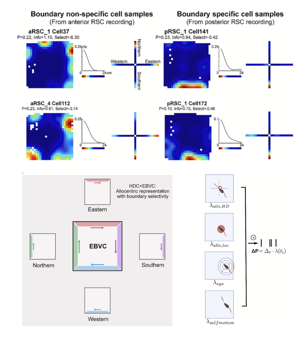
This study reveals a functional variance in the retrosplenial cortex (RSC) for spatial processing. Using calcium imaging in mice, researchers found the anterior RSC (aRSC) predominantly encodes egocentric boundary vectors. In contrast, the posterior RSC (pRSC) integrates this egocentric information with head-direction to create sharpened allocentric boundary representations. Crucially, the function of a specific neuronal subpopulation projecting to the medial entorhinal cortex (RSC→MEC) was identified. These cells form more abstract, global allocentric representations, enriching spatial information for downstream navigation circuits. This refines hierarchical spatial processing models, highlighting subregion- and projection-specific computations within the RSC. Yang Y, Zhang X, Yuan X, Cai H, Cao Q, Miao C*. PNAS, 123 (31) e2600565123

Single field evolution rule governs the dynamics of representational drift in mouse hippocampal dorsal CA1 region.
Research item

Single field evolution rule governs the dynamics of representational drift in mouse hippocampal dorsal CA1 region.

Hippocampal place codes drift across days, but the mechanisms remain unclear. Longitudinal calcium imaging of dorsal CA1 over 56 days during goal-oriented navigation revealed place fields forming, disappearing, and persisting. Statistical and sequential-learning models identified a Single-Field Evolution Rule (SFER): active fields are more likely to remain active next session; inactive fields less likely. SFER simulations reproduced population-level novelty-related stabilization. Extended models incorporating inter-field interactions and daily fluctuations captured coordinated multi-field evolution, ultimately linking single-field activity to hippocampal population drift. Chen C#, Yao S#, Cheng S, Li A, Yan Y, Zhang X, Liu Y, Wang Y, Cao Q, Miao C*. Advanced Science 13 (8), e09532. (#co-first author; * corresponding author)

Differential Impact of Multiple Sensory Deprivation on Spatial-coding Cells in Medial Entorhinal Cortex.
Research item

Differential Impact of Multiple Sensory Deprivation on Spatial-coding Cells in Medial Entorhinal Cortex.

Spatial navigation requires internal maps to remain anchored to external sensory cues. The medial entorhinal cortex (MEC) contains spatially modulated cell types, but how visual and tactile cues jointly support them is unclear. Here we show, using miniature two-photon calcium imaging in freely moving male mice, that sensory deprivation has context-dependent effects. In a visually dominated environment, darkness disrupted all spatially modulated cell types, especially grid and non-grid spatial representations; subsequent whisker trimming had limited additional effects, but more clearly altered headdirection and border-cell tuning. In darkness with salient tactile landmarks, all spatially modulated cell types formed cue-anchored representations, and whisker trimming more strongly impaired grid and spatial coding. Some neurons remained stable after combined deprivation, consistent with support from boundary-related tactile signals. Additionally, a subset of MEC neurons showed activity correlated with whisker movement. These findings reveal flexible integration of visual and tactile information in MEC spatial coding. Tian J, Wen S, Zhou Y, Yao S, Zhang X, Miao C*. Nature Communications. Online publication.

Retrospective grid cells in the medial entorhinal cortex encode past paths during spatial navigation
Research item

Retrospective grid cells in the medial entorhinal cortex encode past paths during spatial navigation

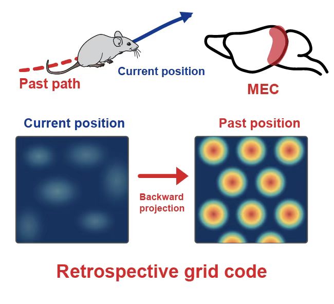
Neurons dynamically switch between prospective and retrospective coding depending on task demands. Prospective coding optimizes future actions, while retrospective coding helps learn from past experiences. The grid cells in the medial entorhinal cortex (MEC) play a key role in spatial navigation, with periodic firing fields to encode current position. While their role in prospective coding has been reported recently, whether and how grid cells exhibit retrospective coding (encoding past paths) remains unclear. Here, we combined large-scale two-photon imaging in freely moving mice with well-established analytical approaches to identify and characterize retrospective grid cells in the MEC. We identified the retrospective grid cells, which encoded path history, showing maximal gridness score at backward-shifted positions (∼15 cm), independent of calcium transient kinetics. Retrospective grid coding showed reduced spatial precision in darkness and was disrupted during assisted movement, indicating that its stable expression depends on sensory and behavioral conditions. In simulations, incorporating retrospective information into a path-integration model reduced accumulated position error and improved model performance across varying movement speeds. These findings support a dual-coding framework where retrospective and prospective cells jointly optimize navigation accuracy. Liu R, Yuan X, He Z, Jin H, Tian J, Chen C, Liu Y, Zhang X, Wen S, Kropff E, Miao C*. Advanced Science. Online publication.

Aging-related cellular and molecular changes reveal a role for Bglap3+ neurons in MEC spatial cognition
Research item

Aging-related cellular and molecular changes reveal a role for Bglap3+ neurons in MEC spatial cognition

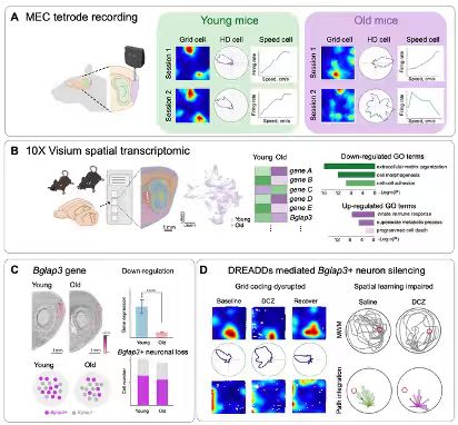
Aging-related cognitive decline, including impairments in spatial orientation and memory, has been linked to dysfunction within the hippocampus and medial entorhinal cortex (MEC). However, the cellular and molecular alterations underlying MEC aging remain poorly understood. Here, we show that old mice exhibit a reduced proportion of grid cells and decreased spatial stability. Transcriptomic profiling of the MEC using 10x Genomics Visium technology reveals 1664 differentially expressed genes between young and old mice. Among these, Bglap3 is identified as a marker gene for a subpopulation of layer III neurons that showed an age-associated reduction in cell number. Chemogenetic silencing of Bglap3+ neurons in young mice impairs spatial tuning of MEC neurons. Together, these findings elucidate cellular and molecular mechanisms contributing to age-related MEC dysfunction and suggest that the loss of Bglap3+ neurons may underlie spatial coding deficits in aging. Cao Q, Suri G*, Cheng S, Liu Y, Liu J, Wen S, Zhong M, Hao J, Yao S, Yang Y, Xu S, Jin H, Yan X, Guo W, Miao C*. Nature Communications. Accepted for publication.

Retrosplenial Cortex Exhibits Parallel Directional Representations and Contributes to Medial Entorhinal Spatial Coding in 3D Curved-Surface Navigation
Research item

Retrosplenial Cortex Exhibits Parallel Directional Representations and Contributes to Medial Entorhinal Spatial Coding in 3D Curved-Surface Navigation

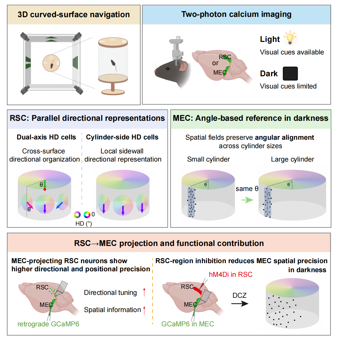
Spatial coding has been extensively studied in two-dimensional (2D) environments, yet how cortical navigation circuits organize directional and positional representations across continuous three-dimensional (3D) curved surfaces remains unclear. Here, using two-photon calcium imaging in freely moving mice exploring a cylindrical arena, we found that the retrosplenial cortex (RSC) exhibited two distinct directional representations. One population showed a cross-surface organization, with preferred directions that shifted systematically as mice moved around the sidewall while remaining coherent with directional tuning on the top platform; this organization persisted in darkness, albeit with reduced precision. A second population exhibited a local sidewall directional representation, with preferred directions that remained stable across sidewall positions and were largely preserved in darkness. Compared with the RSC, the medial entorhinal cortex (MEC) contained fewer and less stable direction-tuned neurons, but showed more precise positional coding. Within the RSC, MEC-projecting neurons carried more precise directional and positional information than the general RSC population. Regional RSC inhibition selectively degraded MEC sidewall positional precision and cross-cylinder angular alignment in darkness, but not in light or in a 2D arena. Together, these findings reveal parallel RSC directional representations and suggest that intact RSC activity contributes to stable MEC positional coding during 3D curved-surface navigation, particularly when visual support is limited. Zhang X#, Yuan X#, Zhang X, Wang Z, Zeng Z, Wang Y, Yang Y, Wen S, Wang S, Xu Q, Tian J, Miao C*. Cell Reports. Accepted for publication.
Multiplexed representation of others in the hippocampal CA1 subfield of female mice
Research item

Multiplexed representation of others in the hippocampal CA1 subfield of female mice

Hippocampal place cells represent the position of a rodent within an environment. In addition, recent experiments show that the CA1 subfield of a passive observer also represents the position of a conspecific performing a spatial task. However, whether this representation is allocentric, egocentric or mixed is less clear. In this study we investigated the representation of others during free behavior and in a task where female mice learned to follow a conspecific for a reward. We found that most cells represent the position of others relative to self-position (social-vector cells) rather than to the environment, with a prevalence of purely egocentric coding modulated by context and mouse identity. Learning of a pursuit task improved the tuning of social-vector cells, but their number remained invariant. Collectively, our results suggest that the hippocampus flexibly codes the position of others in multiple coordinate systems, albeit favoring the self as a reference point. Zhang X, Cao Q, Gao K, Li A, Chen C, Cheng SH, Liu R, H J, Kropff E*, Miao C*. Nature Communications. 15, 3702.